メインコンテンツへスキップ

基礎科学(ハブ)

From Fundamental Sciences to Clinical Translation

fundamental-sciences divider-fundamental

Experimental Outline

einstein. divider-fundamental
  • histology

    Histology

    Histological Sections • Cellular Architecture • Tissue Response

    View Histology

  • chemistry

    Chemistry

    pKa Modulation • Aromatic Compounds • Chemical Stability

    View Chemistry

  • mecanism-action

    Mechanism of Action

    Integrated Biological Response • Metabolic Pathways

    View Mechanism of Action

  • molecular-biology

    Molecular Biology

    Intracellular Signaling • Molecular Regulation

    View Molecular Biology

  • physiology

    Physiology

    Neuromuscular Physiology • Functional Anatomy

    View Physiology

  • lab of anatomy

    Cadaveric Study

    Anatomical Correlation • Human Cadaveric Analysis

    View Study

続きを読む

組織学

Histology

On Wistar rats

Courtesy of Lab of Pathology Department on Medicine School of Ribeirão Preto – USP

histology

01

Study
Group

Experimentators : R.Mené,M.Kim,A.Tenenbaum

N= 29 Wistar rats
Weight mean : 421g

Origin of animals : Biotério Central of Campus of Ribeirão Preto of São Paulo University
Maintenance : Experimental Pathology Lab of Pathology Department on Medicine School of Ribeirão Preto – USP
Animals keeping : polypropylen boxes
Animals food : basic diet of lab and water
Light conditions : natural light/dark cycles under controlled basic conditions
Experimentators : A.Tenenbaum,R.Mené,Kim

pathology department

02

Animals Sacrifice & Histologic Samples

Conventional Microscopy

The mice were euthanized in CO2 chambers.

  • Incision of the skin with a blade nº 23
  • Finding and isolation of the pretibial muscle using Metzenbaum scissors
  • Section of tendons with a Mayo scissors.
  • Fixation of Portions of the muscles on sections of cork oak
  • Embedding using 50% formaldehyde solution - 10% buffered and 50% of pure paraffin for conventional optical microscopic study.
  • Staining Sections as routine histological process.
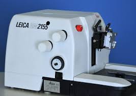
  • Microtome used : Leika RM 2155
  • Coloration of muscle slices with

-hematoxilin-eosin
-Masson´s trichrome
-and red picro-sirius.

  • Histologic examination was realized with a conventional microscope (Olympus BH-2).
  • Observation under polarized lampe
  • Photographic documentation was realized with a Leika DMR microscope joined to a digital camera Leika CD 300F and compatible PC.
microtome

03

Material

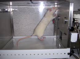
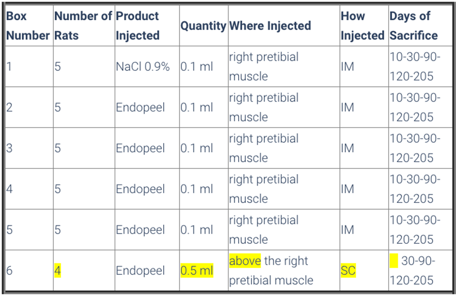
6 rats/box

  • Animals were divided in 6 boxes, so that boxes from number 1 to 5 had 5 rats and box number 6 had 4 rats 
  • All animals were submitted to a walking evaluation by footprint impression on special paper , and previous immersion of back legs on a soap solution. 
  • Box 1 had the control group, that received the application of 0,1 ml saline solution ( NaCl 0,9%) on the right pretibial muscle. 
  • The mice of boxes 2 to 5 received 0,1 ml of Endopeels Original Main Product on the same muscle as control group.

  • Box 6 was constituted of 4 rats, and each one received 0,5 ml of Endopeels original main product injected on the subcutaneous layer. 

  • All the animals were submitted to walking evaluation before and after their respective injections.

boxes for rats

04

Methods

Animals are sacrified after 10 days-1 month- 3 months-7 months

sacrifice table

05

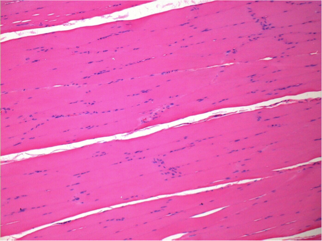
Control

After injection of NaCl 0.5%

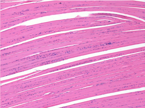
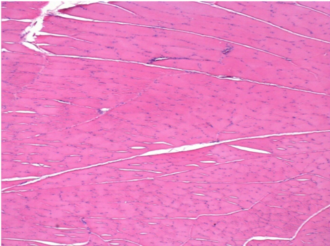
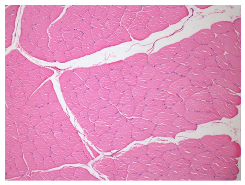
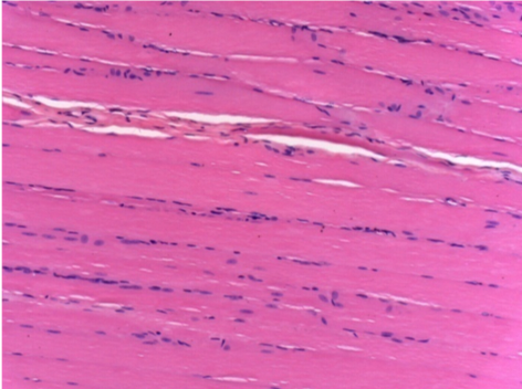
Comment : Nothing to declare after saline solution injection

without treatment

L:Pretibial-No treatment

R:Pretibial-After 0.1 ml NaCl 0.9% IM

06

10 days after Endopeel Injection

Box 2

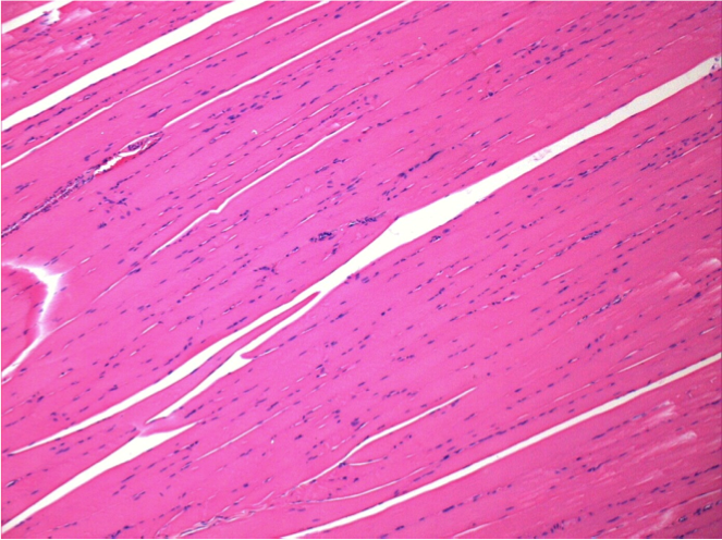
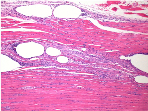
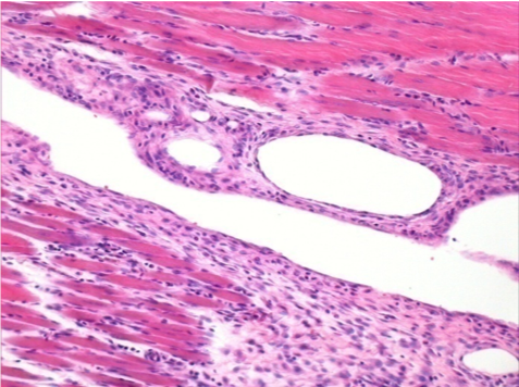
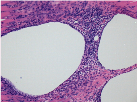
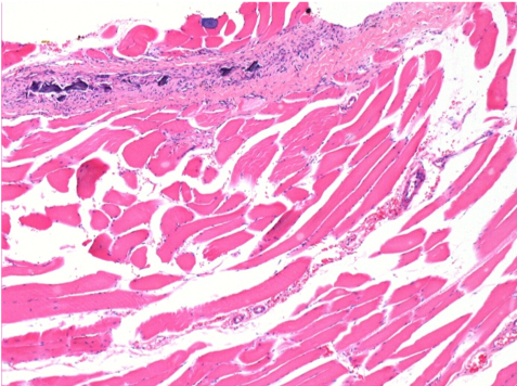
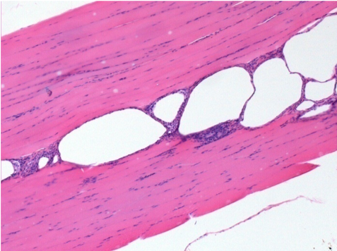
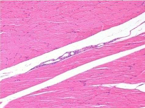
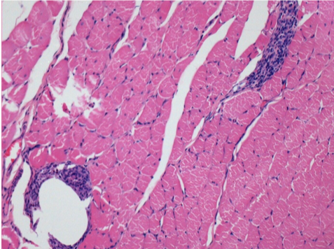
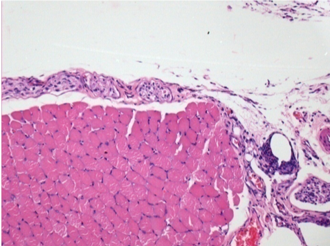
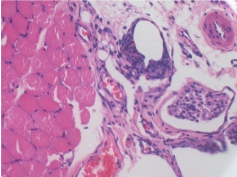
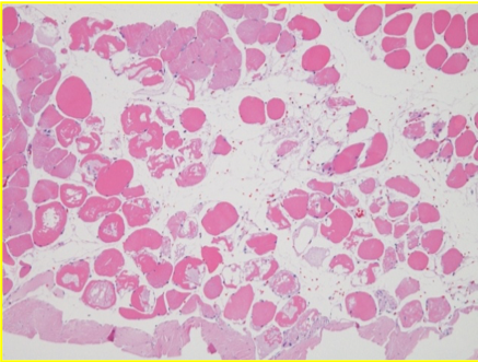
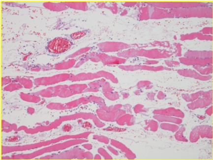
10 days after Endopeel Injection 0.1ml in the right pretibial muscle.

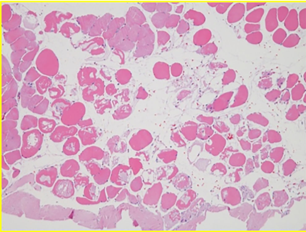
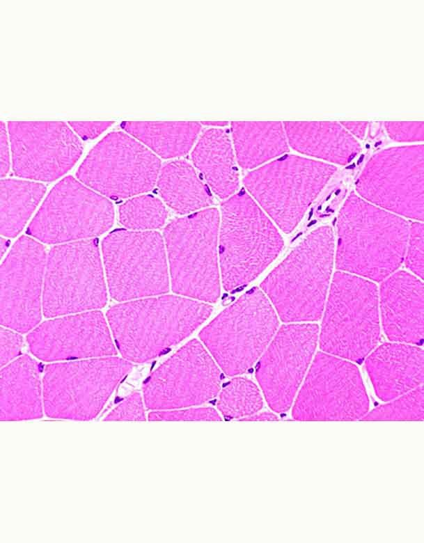
Here you may see the formation of the vacuoles which are surrounded by lymphocytes. Vacuoles are different from tissue necrosis . The presence of lymphocytes is related to the permeability of the cell membranes.


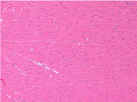
left control- 100xD10

L : Control-100xD10

right 100xD10

R:100xD10

right200xx

R :200xD10

right400xxD10

R :400xD10

07

1 month after Endopeel Injection

Box 3

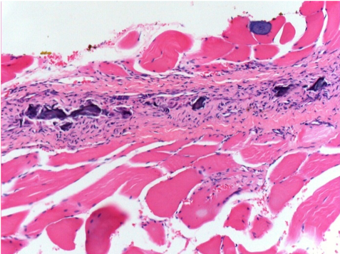
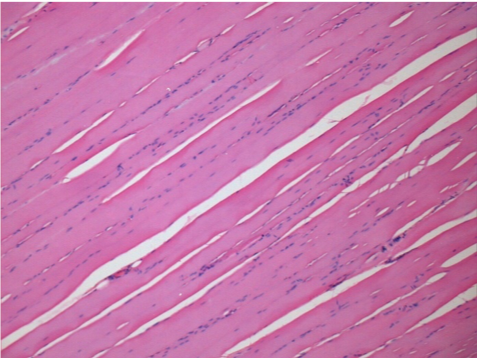
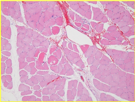
1 month after Endopeel Injection 0.1ml in the right pretibial muscle.
What is seen in black on the pictures is not a necrosis like could imagine some scientifics !
In fact, 4 conclusions have to be taken in consideration

  • an artefact of coloration
  • an absence of necrosis
  • an apoptosis
  • a bioregenerative process 

Read more about the bioregenerative process

left control- 100xD30

L : Control-100xD30

right 100x

R:100xD30

Necrosis Definition

right200xxD30

R :200xD30

right400xxD30

R :400xD30

08

3 months after Endopeel Injection

Box 4

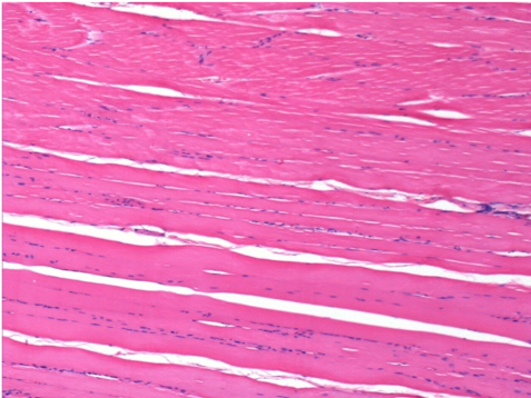
3 months (D90)after Endopeel Injection 0.1ml in the right pretibial muscle.

left control- 100xD90

L : Control-100xD90

right 100x

R:100xD90

09

7 months after Endopeel Injection

Box 5

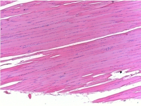
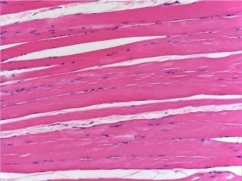
7 months (D210)after Endopeel IM Injection 0.1ml in the right pretibial muscle.

Complete Restitutio ad integrum after 7 months

left control- 100xD210

L : Control-100xD210

right 100x

R:100xD210

L Control 50xD210

L :Control 50xD210

R50X-D210

R50X-D210


10

Endopeel Injection in Subcutaneous Tissue

Box 6

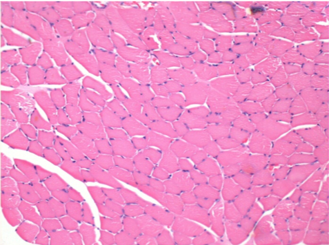
0.5 ml ( 5x 0.1ml)  Endopeel SC Injection in the right subcutaneous pretibial area.

L:200x-Control-SC

R-D10-SC-200X

R-D10-SC-200X

R-D30-SC-200X

R-D30-SC-200X

R-D90-SC-200X

R-D90-SC-200X

R-D210-SC-200X

R-D210-SC-200X

R-D210-SC-400X

R-D210-SC-400X


conclusions
    • Endopeel induces a selective reversible myofibrolysis and  inflammatory reaction on a period of 1 month, approximately
    • Muscular changes are reversible in almost   full totality
    • The muscle is the better place to inject Endopeel because of more efficacity, control and duration of  its action
    • No necrosis nor abcess have been found all over the study.

Other Study applied to Gynecology

Read More

続きを読む

アポトーシス

Apoptosis

Definition

  • in line with the body's metabolic and homeostasis plan

    for the defense and prevention of genetic mutations or viral attacks,which endanger the  survival of the organism

  • Apoptosis is a selective mechanism subjected to a regulatory plan of survival and death factors

    When DNA damage occurs,the factors are activated by the environment surrounding the damaged cell,as information from other nearby cells,which they receive from the damaged cell,the information needed to prepare the rescue plan.The signaling of survival factors reach the damaged cell,activating the genetic resident control factor program.In DNA transcriptional and regulatory factors attempt rescue,seeking to repair the damage.Programmed cell death factors are activated via caspases,which are proteins that form a scaffold at the level of the cytosolic plasma membrane,and they act by activating a proteolytic cascade regulated by phosphorylations.Mitochondrial cytochrome c,released into cytosol,participates in activation of caspases.

  • Apoptosis may be triggered by an external signal

    Phosphatidylserine moves from the position inherent in the cytosolic plasma membrane,going outside on the extracellular plasma membrane,and it is formed as if it were a receptor with external signaling factor on the cell in view of the macrophages,which intercept the signal positioning themselves on the cell,then incorporate it and degrade it in their body.


Main causes leading to programmed cell death

  • Oxidative stress

    caused by signaling factors that come from the external environment(magnetic radiation,reactive chemical compounds and gases).

  • Reactive factors

    such as Ros(highly reactive products of the partial reduction of O2,superoxide *O-2,minor byproduct of oxidative phosphorylation)

  • Viral infections or stress

    such as heat shock


Bioenergetic signaling mechanism of apoptosis

  • Signaling is information in the form of energy in living beings

    by animating metabolic cycles and therefore matter.

    Through chemical-biological evolution,the cell has managed to develop a constitutive plan,animated by complex energy processes,where matter is transformed through energy by regulating and making the metabolic systems of living beings function.Matter to be animated by energy,there must be an internal order in living beings,which needs an energy supply to make it work. This is a thermodynamic plane,which must ensure sufficient availability of energy,which is supplied and then extracted from the matter that animates the surrounding Cosmos.


  • Maintaining a certain type of order in the living organism

    that keeps it away from a equilibrium that can bring it to a steady state,responsible for the decay of the matter.


Apoptosis is made necessary

  • when damage in DNA jeopardizes a certain type of order

    consisting of a stationary-dynamic plane,which keeps away from the dynamic order of reactions

  • The road to old age is littered

    with a constant accumulation of mutations in the DNA,which the subsequent statistical increase in disease and cell senescence,directly proportional to the errors and damages that accumulate in the DNA or inside the macromolecules,regulated by biosynthesis.


Apoptosis & inflammation

  • Apoptosis is an effective means of preventing irreversible mutations

    that add up and are transmitted to the subsequent progeny of cells

  • Arachidonic acid is an essential component of Endopeel

    and this is manipulated in such a way that the entire molecular composition amplifies a cascade of reactions,which reactions initiate a renewal of the tissue that leads to an apoptotic process

  • Endopeel gives the necessary impetus to an energetic process

    that makes the mechanism of survival and death factors more selective.The selective process,on the one hand with survival factors,triggers an attempt to rescue the cell from mutations that could endanger the organism.If this rescue attempt does not work within a set time,phosphatidylserine from the inside of the plasma membrane slides outwards,capturing the signals triggering a caspase cascade ends the life of the cell,which is directed towards apoptosis.


  • Conclusion

    Apoptosis is an essential concept for slowing down the senescence process.

    As it is well described in the article "Journal of Advanced Research".The homeostasis processes between anti-inflammatory and inflammatory factors are directly linked to lifestyle,and therefore aging itself is also an accumulation of mutations.The disease that makes its way first is aging,the general structure suffers irreversible damage.The problem is precisely to arrive in time before these damages are irreversible.For this reason,apoptosis is an essential concept for slowing down the senescence process.

Ageing: Is there a role for arachidonic acid and other bioactive lipids ?

Do you still have questions left?

このメールアドレスはスパムボットから保護されています。閲覧するにはJavaScriptを有効にする必要があります。

続きを読む

壊死

Necrosis

Main Etiologies

Cells that accidentally die in response to acute damage,trauma,lack of blood supply and chemicals/drugs.

These cells are not subjected to any regulatory and selective mechanism,and thus die through a process of cell necrosis

Acute Damage
Trauma
Lack of Blood Supply
Chemicals or Drugs
necrosis hidroxibenceno

Necrosis seen with dangerous copies

25 min post endopeel

          disorganization seen 25 min post endopeel

reorganization 60 min after endopeel

reorganization 60 min after endopeel

EXPERIMENT

after 0.05 ml endopeel injection

Department of  Organ and System Research, Akita University, School of Medicine and YMC-Biyou Yokohama Minato and Shinjuku Clinics

ANALYSIS :Sapporo pathological Research Institute Ltd.

Consequences

Cells in a state of necrosis swell and explode,pouring their contents into the surrounding region,contaminating the environment in which the other cells reside,causing and triggering an inflammatory response,spreading like a chessboard,advancing as a degrading factor.

Necrosis is a process in which there is a loss of energy (therefore loss of signaling factors),leading to defects in the metabolism consequent to the loss of the ionic gradients(these gradients present on the 2 sides of the plasma cell membrane.)

続きを読む