Zum Hauptinhalt springen

Ursprung der Myomodulation

Discovering the Origins of the Myomodulation :
A Revolutionary Approach to Aesthetics

We don’t follow trends — we build what’s next.
Merging vision and code, we turn bold ideas into digital reality.
Two minds, one mission: redefining what a web agency can be.


mauro-tiziani stemma-zaguarina

Co-Inventor & Graphic Designer

Mauro Tiziani is the co-inventor of the Myomodulation and the creative mind behind its visual identity.
As a graphic designer and developer, he transforms complex medical principles into clear, dynamic visuals that educate both professionals and patients.
He crafted the entire visual ecosystem of Myomodulation— from 3D models and apps to educational media — ensuring that science remains understandable and aesthetically engaging.


dr-alain-tenenbaum stemma-pulzinetto

Medical Strategist & Webmaster

Dr Alain Tenenbaum is the scientific architect of the Myomodulation.
A pioneer in aesthetic medicine and digital education, he developed the medical, biochemical and strategic frameworks that support the method.
Founder and administrator of over 30 specialized medical portals, he uses the web as a scientific and educational tool — bridging research, publication, and training worldwide.

Platforms • Protocols • Training

corniche-piumette.

Why is this duo unique

While most physicians outsource their web and design work, they keep everything in-house — mastering both the medical and the digital sides of innovation.
This rare combination ensures full control, authenticity, and coherence between science and visual identity.

corniche-piumette

Our Values & Philosophy

We believe in purpose-driven design and future-proof technology.
Our clients aren't just customers — they're collaborators, co-creators, and the heart of every solution.
Our philosophy: think big, aim higher, and turn bold ambition into digital success.
Weiterlesen

Bioregeneration bei Myomodulation

Bioregenerative

Myomodulation® provides a revolutionary approach to treat the aging appearance and organs ( skin, muscles) that will provide patients with more long-lasting and youthful results.

       Services Provided

  • Rejuvenation

  • Embellishment

  • Tissue Reshaping

To Resume

  • 3D Isovolumetric Tissular repositioning

  • Myoplasty

  • Myopexy

  • Myotension

  • Bioregeneration

The skin of the face is one of the areas most affected by the passage of time. It is even possible that it suffers from phenomena of premature aging if we do not take care of it properly. Crow's feet, sagging in the mouth and cheekbones, the bar-code effect, forehead wrinkles, all these effects and more are symptoms of aging of our skin. There are many ways to combat these phenomena, including MYOMODULATION techniques.

There is indeed a large amount of medico-aesthetic treatments to improve the appearance of the skin of the face and to find a younger appearance. There are also many treatments that can be associated with each other for an ever more effective result.

Weiterlesen

Wirkungsmechanismen

Mechanistic Overview

Mechanisms of Action

Endopeel® acts through a combination of immediate physicochemical interactions and longer-lasting functional tissue effects. Its clinical activity is not limited to simple filling or swelling, but involves structural tissue interaction, selective response of hypotonic compartments and progressive functional support of treated anatomical areas.

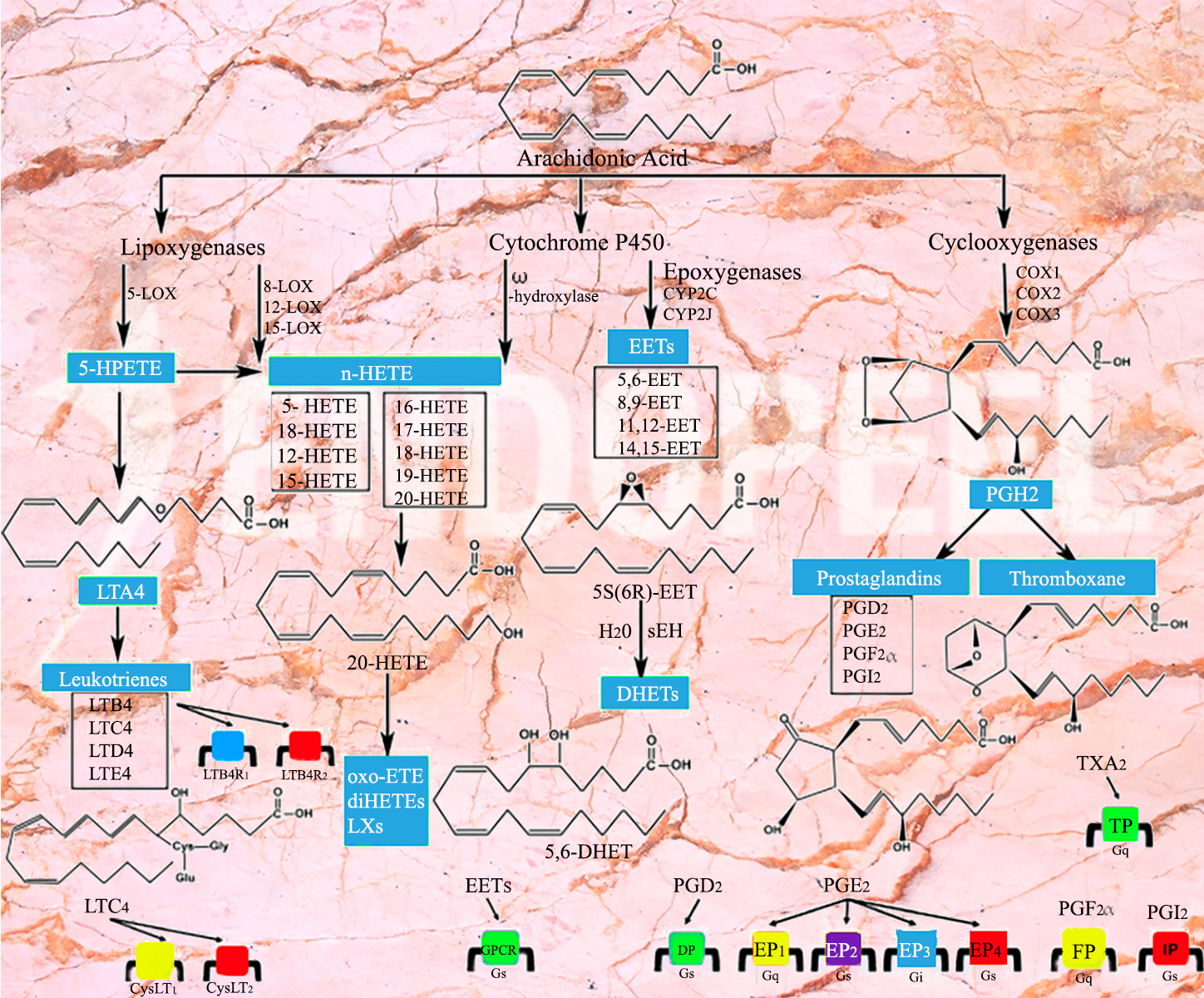
Arachidonic acid cascade illustrating molecular pathways potentially involved in Endopeel mechanisms of action
Figure 1. Biochemical pathways related to the arachidonic acid cascade illustrating molecular interactions potentially involved in the mechanisms of action of Endopeel® at the cellular level.

Quick Scientific Overview

Immediate Tissue Interaction

The first phase involves local physicochemical interaction with hypotonic tissues, contributing to structural densification and an immediate support effect.

Functional Support

Endopeel® contributes to functional myostructural support rather than simple volumization, helping reposition and reinforce selected soft-tissue areas.

Progressive Biological Effects

Longer-term observations suggest structural and neurobiological responses that may help explain the sustained clinical duration seen in selected indications.

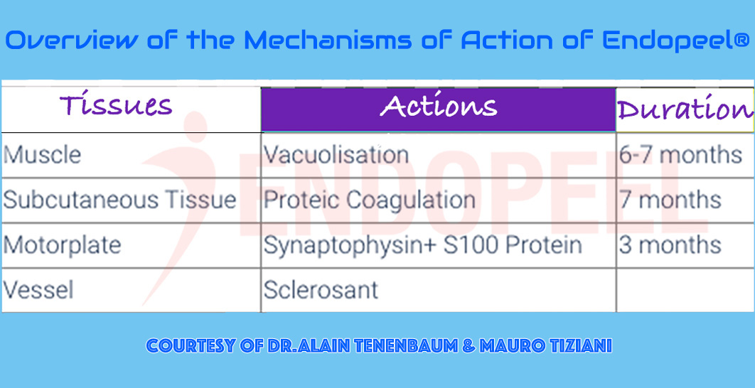
Overview diagram summarizing the proposed mechanisms of action of Endopeel at molecular and tissue levels
Figure 2. Schematic overview summarizing the proposed mechanisms of action of Endopeel®, integrating physicochemical interactions, microstructural tissue modifications and functional support effects observed in hypotonic tissue compartments.

Molecular and Tissue-Level Activity

Endopeel® should be understood as a procedure with both physicochemical and functional consequences. At tissue level, the injected product interacts with selected planes showing hypotonia, laxity or poor structural support. This interaction may explain the immediate change in firmness observed in certain areas.

At the same time, the clinical effect appears to go beyond a transient reaction. Experimental and clinical observations suggest that structural stabilization, tissue support and specific biological responses may contribute to the persistence of results over time.

  • Immediate physicochemical tissue response
  • Selective action in weak or hypotonic tissue environments
  • Progressive structural support rather than simple filling
  • Potential biological contribution to prolonged clinical effect
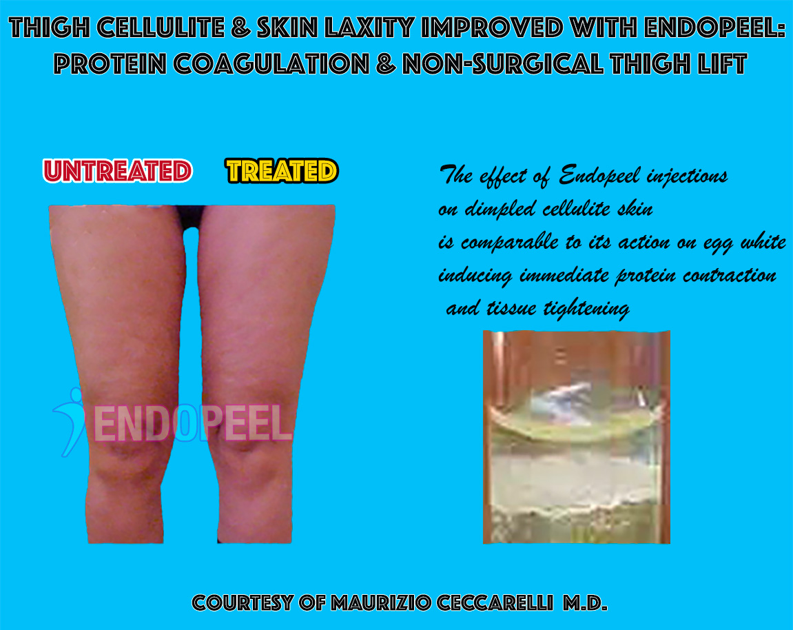
Clinical example of proteic coagulation in hypotonic subcutaneous tissues
Figure 3. Clinical illustration of cellulite treatment showing the visible effects of Endopeel® on hypotonic subcutaneous tissues. Local physicochemical interaction may contribute to improved structural support and contour stabilization.
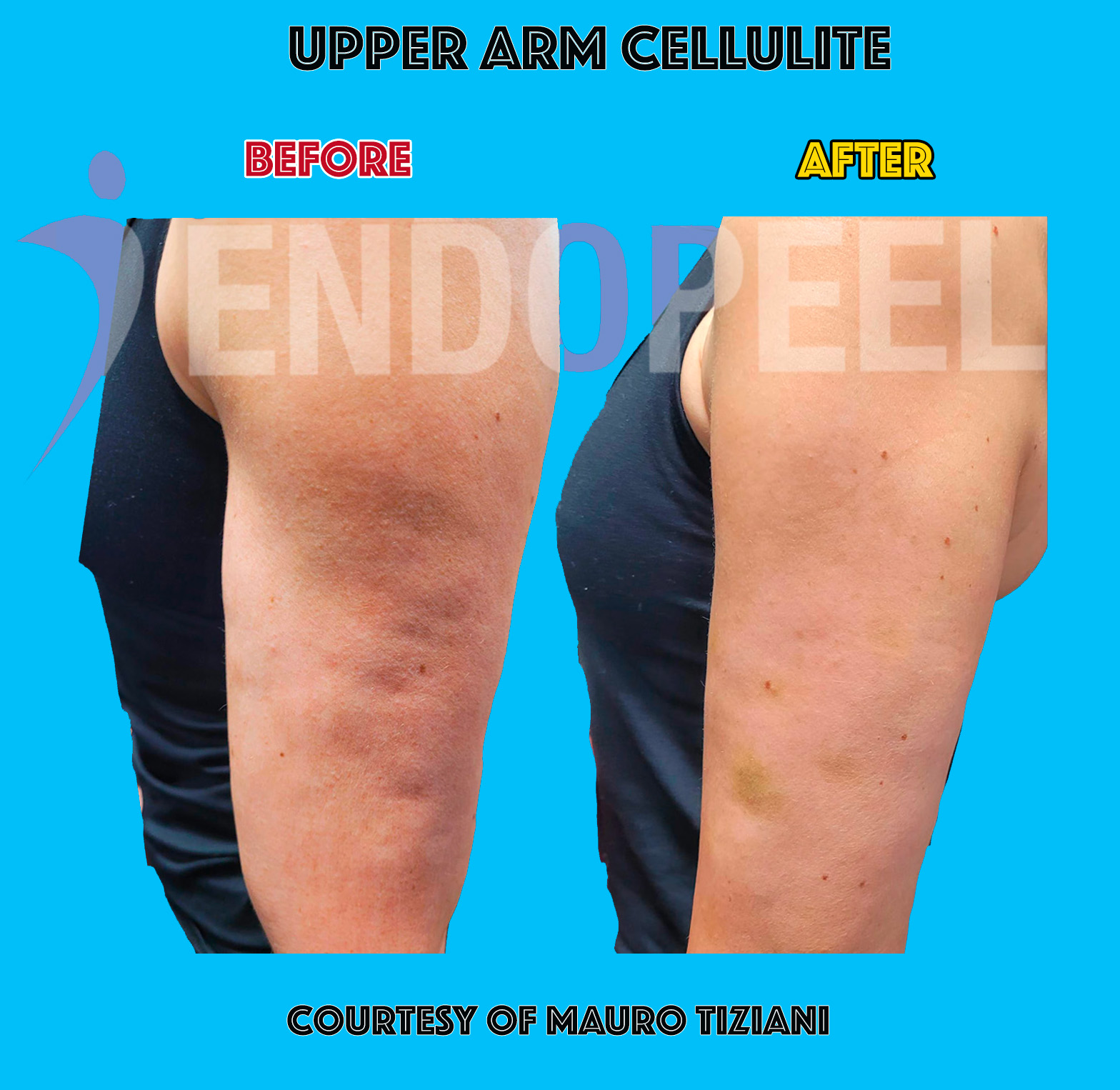
Endopeel treatment of cellulite demonstrating tissue structural support
Figure 4. Clinical example of tissue response following Endopeel® injection in areas affected by hypotonicity and structural weakness. The treatment may contribute to functional tissue reinforcement and improved mechanical behavior of the treated plane.
Core Mechanism

Proteic Coagulation of Hypotonic Subcutaneous Tissues

One of the proposed immediate mechanisms is a selective proteic coagulation effect in hypotonic subcutaneous tissues. In practical terms, this may produce local densification and better mechanical support in tissue compartments that have lost firmness.


This helps explain why the clinical effect is often perceived as tightening, repositioning or lifting rather than as simple volumetric augmentation.


The response appears especially relevant in areas where tissue laxity and structural weakness are central components of the deformity.


Clinical interpretation: the effect is better described as functional tissue reinforcement with visible contour support.
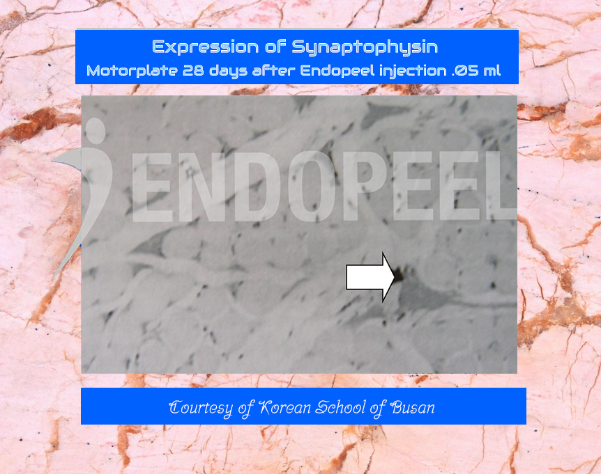
Synaptophysin-related experimental observation after Endopeel treatment
Figure 5. Experimental observation suggesting synaptophysin expression in treated tissue areas. Synaptophysin is commonly used as a marker of synaptic vesicles and neuronal activity, suggesting that Endopeel® may interact not only with tissue structure but also with elements related to neuromuscular function.

Synaptophysin and Neurobiological Observations

Experimental observations have suggested the involvement of synaptophysin-related changes after Endopeel® treatment. Synaptophysin is commonly regarded as a marker associated with synaptic vesicles and neuronal activity, making such findings particularly interesting from a mechanistic perspective.


These findings indicate that the procedure may not be limited to a passive structural effect. Instead, the treatment could interact with biological pathways related to neuromuscular signaling and local functional behavior of the treated tissues.


Although additional research is needed to fully clarify these mechanisms, such observations contribute to the hypothesis that Endopeel® may act as a form of functional myoplasty, influencing both tissue mechanics and neuromuscular dynamics.

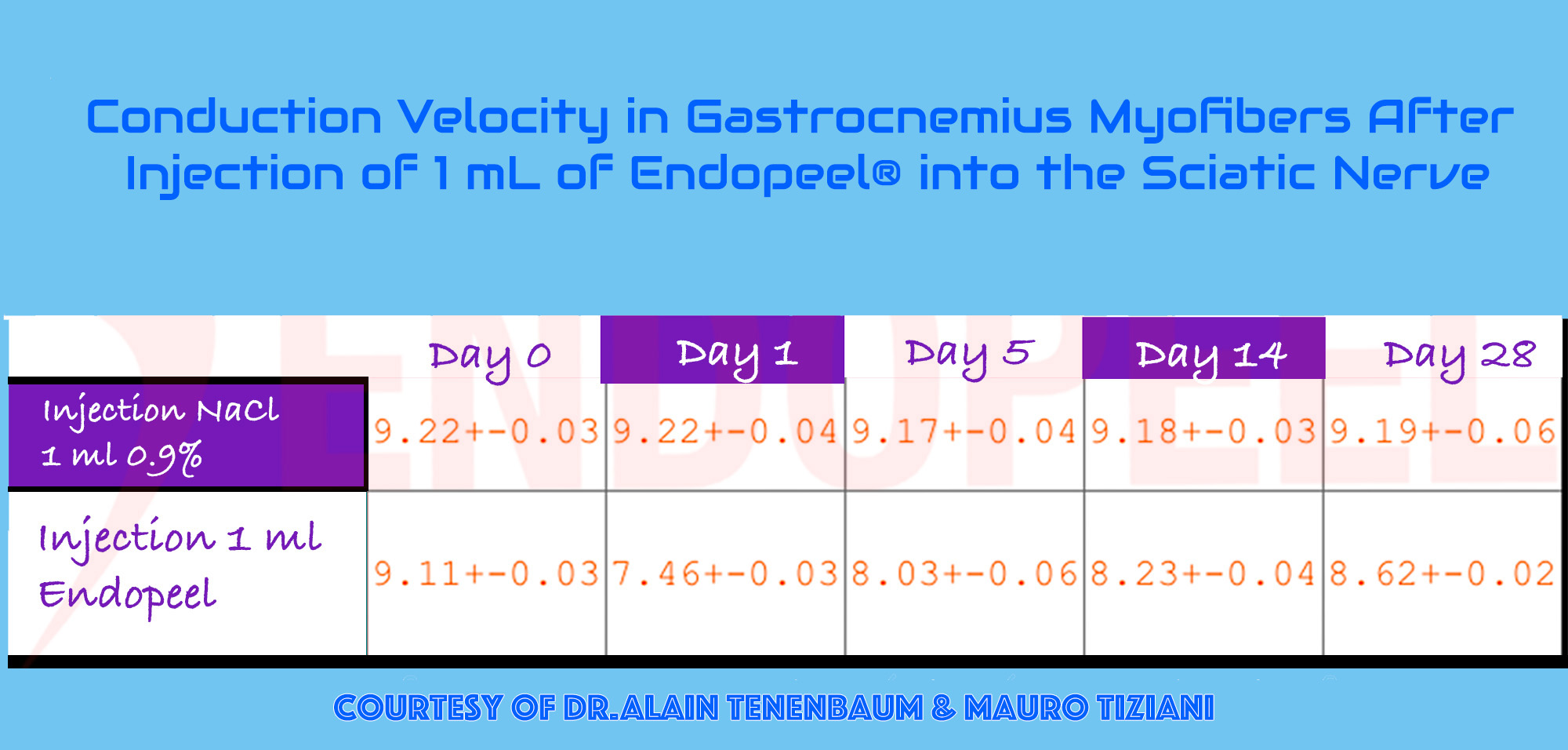
Conduction velocity observations related to Endopeel functional myostructural support
Figure 6. Experimental observations related to conduction velocity in treated tissues, suggesting that Endopeel® may influence not only structural support but also functional behavior within selected myofibers and adjacent support planes.

Conduction Velocity and Functional Myostructural Support

Another important area of investigation concerns conduction-related observations within treated myofibers and adjacent support structures. These findings are consistent with the idea that Endopeel® may influence not only tissue consistency but also functional behavior in selected muscular territories.


This is one of the reasons the technique has been positioned as a method of functional myoplasty or myopexy. The objective is not to artificially occupy space, but to help restore support, tension balance and contour through selective structural action.


  • Functional tissue support
  • Possible interaction with conduction behavior
  • Reinforcement of weak structural planes
  • Improved contour through support, not overfilling

Vascular and Periorbital Effects

In selected indications such as dark circles, the visible improvement may involve more than simple camouflage. Better support of the local tissue environment may influence the appearance of vascular show-through, shadowing and periorbital depression.


Periorbital dark circles are known to have a multifactorial origin including vascular show-through, tear trough depression, shadowing effects and, in some cases, pigmentary components. Structural support of the periorbital tissue may therefore modify the optical appearance of the region by reducing shadowing and improving light reflection across the lower eyelid contour.


The effect on vessels should therefore be interpreted cautiously and in context: not as a classic vascular treatment, but as a structural intervention that can modify how the overlying tissues behave and how vascular coloration becomes clinically visible.

Periorbital implication: improvement may result from better support, reduced transparency effect and modified light reflection.
Clinical improvement of dark circles
Figure 7. Clinical example of periorbital improvement. Structural support of the local tissue environment may reduce the visible appearance of vascular show-through and shadowing, contributing to a more homogeneous periorbital contour.
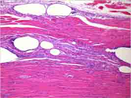
Histological section showing vacuolization phenomena after Endopeel injection
Figure 8. Histological section illustrating vacuolization phenomena observed in treated tissue planes after Endopeel® injection. Such microstructural changes may reflect localized physicochemical interactions within the tissue environment and a transient rearrangement of tissue architecture.

Histology, Microstructure and Experimental Evidence

Histological observations following Endopeel® injections have revealed areas of localized vacuolization and microstructural modification within the treated tissue planes. These findings suggest that the procedure may induce a physicochemical interaction with the surrounding tissue environment rather than producing a purely superficial cosmetic effect.


The presence of vacuolization may represent a transient reorganization of the microstructure of the tissue. Such changes could influence local mechanical properties, including tension distribution and structural behavior of hypotonic tissue compartments.


When these histological observations are considered together with clinical outcomes, they contribute to a plausible biological framework linking localized tissue interaction, microstructural rearrangement and the functional support effects observed in practice.

Additional microscopic material and detailed histopathological observations are available in the dedicated histology section.


See detailed histopathological material →

Duration of Action

The duration of Endopeel® effects may vary according to the indication, tissue quality, technique and functional loading of the treated area. Clinical and observational data nevertheless suggest that some effects are not merely immediate but may persist over several months depending on the treated anatomy.

Immediate
Structural support effect may appear rapidly after injection.
Short-term
Functional improvement may continue to stabilize during the first weeks.
Mid-term
Several clinical observations indicate persistence over a period of months.
Variable
Duration depends on anatomy, indication, technique and maintenance strategy.
Figure 9. Conceptual timeline illustrating the typical progression of clinical effects observed after Endopeel® treatment. Immediate structural interaction may be followed by functional stabilization and persistence of clinical outcomes over several months depending on the anatomical indication and tissue characteristics.

Why These Mechanisms Matter Clinically

Understanding the mechanisms of action is essential to explain why Endopeel® should not be confused with classic fillers, simple volumizing procedures or purely superficial correction techniques.


The proposed mechanisms suggest a treatment rationale based on structural tissue interaction, functional support and selective reinforcement of hypotonic compartments. This framework helps explain the clinical relevance of the technique in indications characterized by tissue laxity, support failure and biomechanical instability.

Mechanistic synthesis

Taken together, these observations suggest that Endopeel® may act through a combination of localized physicochemical interaction, microstructural tissue reorganization and functional support of hypotonic anatomical compartments.


The integrated framework helps explain the clinical effects observed across different indications while remaining compatible with the experimental and histological findings currently available.

  • Functional myoplasty concept
  • Structural support rather than volumization
  • Selective interaction with hypotonic tissues
  • Coherent mechanistic framework
Figure 10. Conceptual summary of the clinical interpretation of Endopeel® mechanisms of action, integrating physicochemical tissue interaction, microstructural modification and functional support effects observed in clinical practice.
Scientific note

The mechanisms described above represent an interpretative framework based on clinical observations, experimental material and histological findings reported in the Endopeel® literature. As with many minimally invasive techniques, several biological aspects remain the subject of ongoing investigation and should be interpreted within the context of currently available evidence.

Weiterlesen