Mechanisms of Action

Mechanistic Overview

Mechanisms of Action

Endopeel® acts through a combination of immediate physicochemical interactions and longer-lasting functional tissue effects. Its clinical activity is not limited to simple filling or swelling, but involves structural tissue interaction, selective response of hypotonic compartments and progressive functional support of treated anatomical areas.
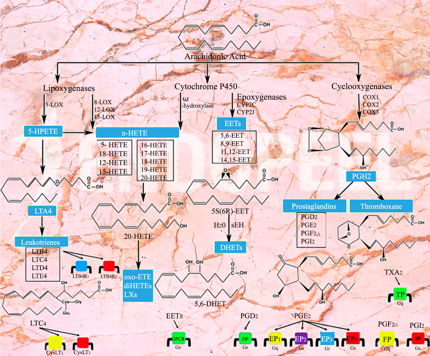
Arachidonic acid cascade illustrating molecular pathways potentially involved in Endopeel mechanisms of action
Figure 1. Biochemical pathways related to the arachidonic acid cascade illustrating molecular interactions potentially involved in the mechanisms of action of Endopeel® at the cellular level.

Quick Scientific Overview

Immediate Tissue Interaction

The first phase involves local physicochemical interaction with hypotonic tissues, contributing to structural densification and an immediate support effect.

Functional Support

Endopeel® contributes to functional myostructural support rather than simple volumization, helping reposition and reinforce selected soft-tissue areas.

Progressive Biological Effects

Longer-term observations suggest structural and neurobiological responses that may help explain the sustained clinical duration seen in selected indications.
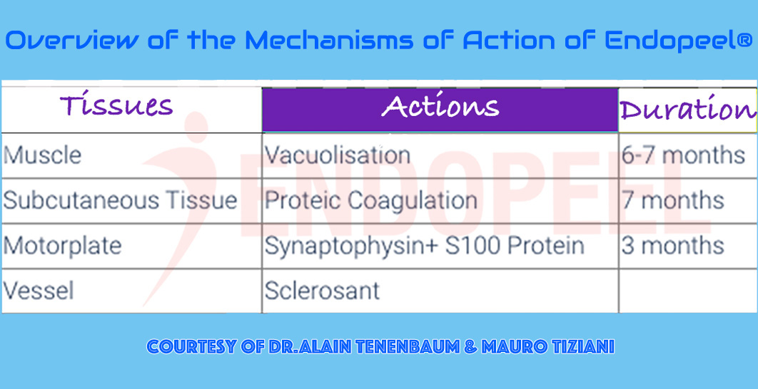
Overview diagram summarizing the proposed mechanisms of action of Endopeel at molecular and tissue levels
Figure 2. Schematic overview summarizing the proposed mechanisms of action of Endopeel®, integrating physicochemical interactions, microstructural tissue modifications and functional support effects observed in hypotonic tissue compartments.

Molecular and Tissue-Level Activity

Endopeel® should be understood as a procedure with both physicochemical and functional consequences. At tissue level, the injected product interacts with selected planes showing hypotonia, laxity or poor structural support. This interaction may explain the immediate change in firmness observed in certain areas.

At the same time, the clinical effect appears to go beyond a transient reaction. Experimental and clinical observations suggest that structural stabilization, tissue support and specific biological responses may contribute to the persistence of results over time.

  • Immediate physicochemical tissue response
  • Selective action in weak or hypotonic tissue environments
  • Progressive structural support rather than simple filling
  • Potential biological contribution to prolonged clinical effect
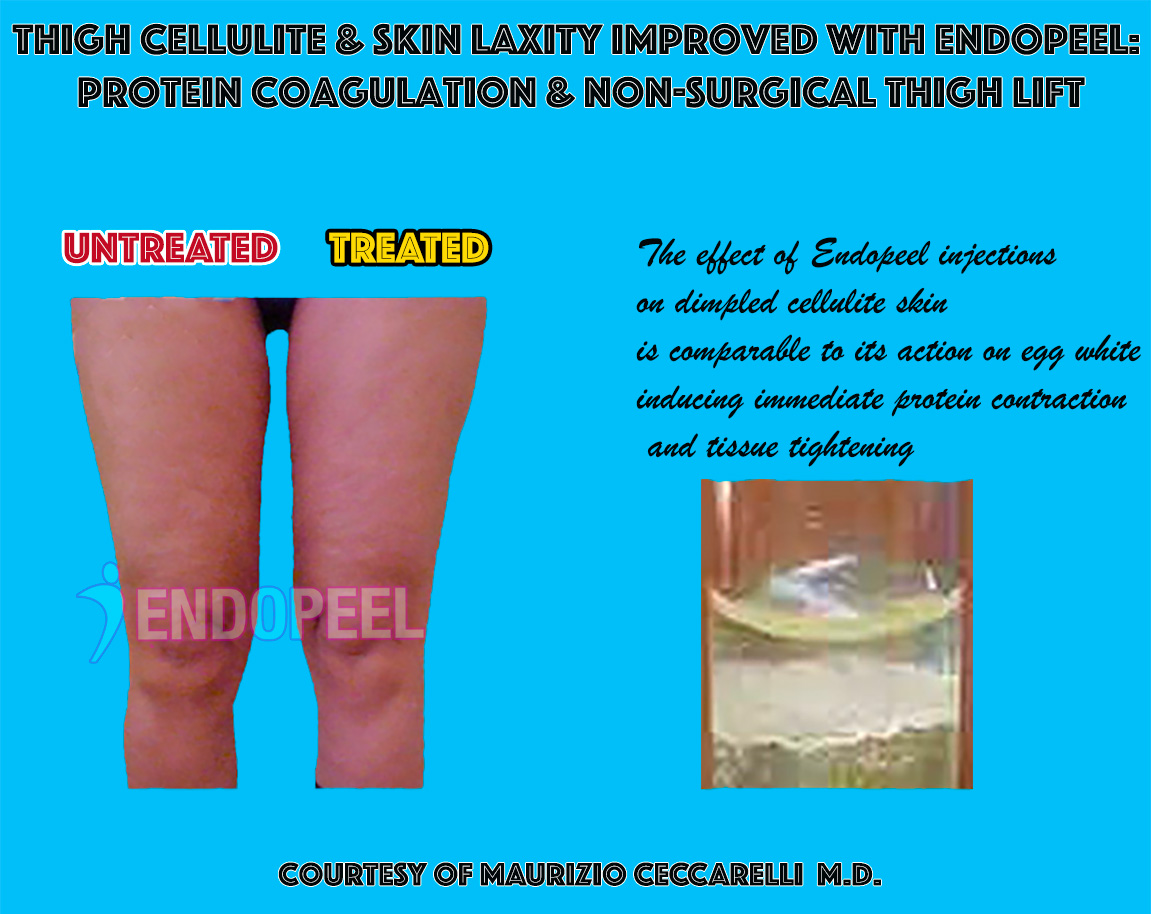
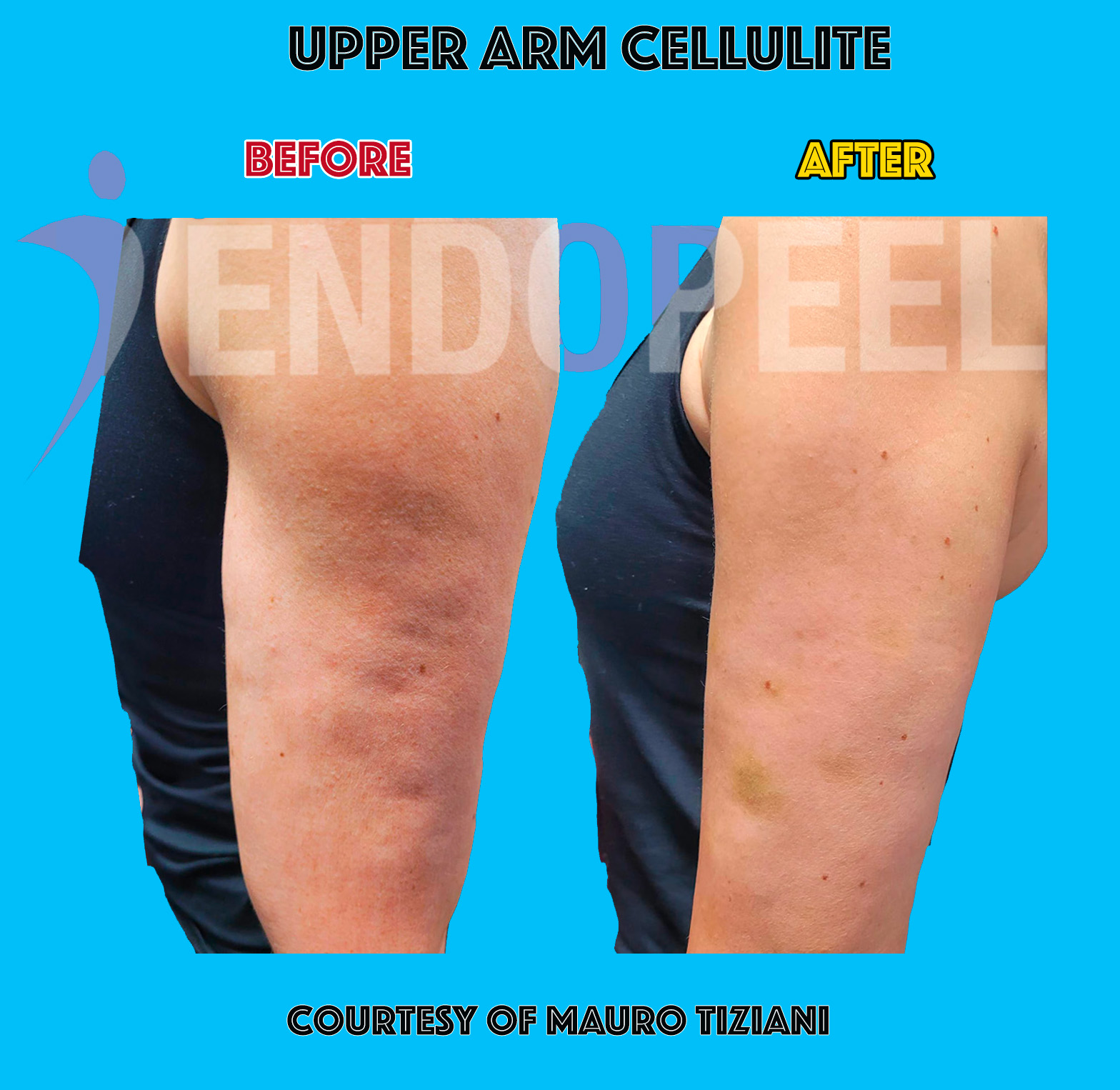
Clinical example of proteic coagulation in hypotonic subcutaneous tissues
Figure 3. Clinical illustration of cellulite treatment showing the visible effects of Endopeel® on hypotonic subcutaneous tissues. Local physicochemical interaction may contribute to improved structural support and contour stabilization.
Endopeel treatment of cellulite demonstrating tissue structural support
Figure 4. Clinical example of tissue response following Endopeel® injection in areas affected by hypotonicity and structural weakness. The treatment may contribute to functional tissue reinforcement and improved mechanical behavior of the treated plane.
Core Mechanism

Proteic Coagulation of Hypotonic Subcutaneous Tissues

One of the proposed immediate mechanisms is a selective proteic coagulation effect in hypotonic subcutaneous tissues. In practical terms, this may produce local densification and better mechanical support in tissue compartments that have lost firmness.


This helps explain why the clinical effect is often perceived as tightening, repositioning or lifting rather than as simple volumetric augmentation.


The response appears especially relevant in areas where tissue laxity and structural weakness are central components of the deformity.

Clinical interpretation: the effect is better described as functional tissue reinforcement with visible contour support.
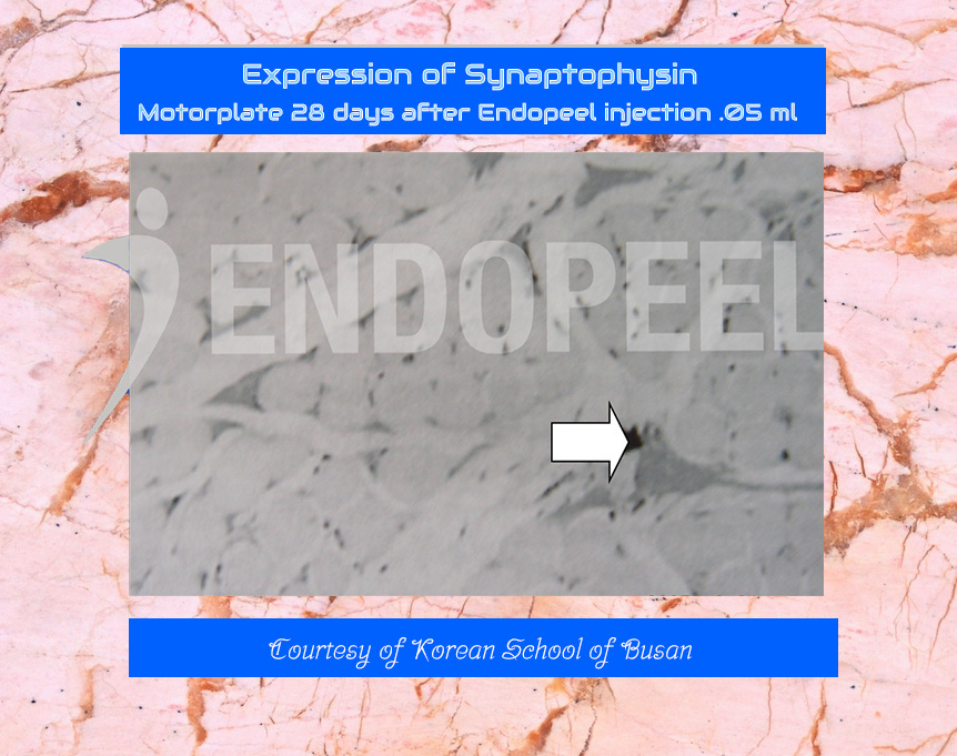
Synaptophysin-related experimental observation after Endopeel treatment
Figure 5. Experimental observation suggesting synaptophysin expression in treated tissue areas. Synaptophysin is commonly used as a marker of synaptic vesicles and neuronal activity, suggesting that Endopeel® may interact not only with tissue structure but also with elements related to neuromuscular function.

Synaptophysin and Neurobiological Observations

Experimental observations have suggested the involvement of synaptophysin-related changes after Endopeel® treatment. Synaptophysin is commonly regarded as a marker associated with synaptic vesicles and neuronal activity, making such findings particularly interesting from a mechanistic perspective.


These findings indicate that the procedure may not be limited to a passive structural effect. Instead, the treatment could interact with biological pathways related to neuromuscular signaling and local functional behavior of the treated tissues.


Although additional research is needed to fully clarify these mechanisms, such observations contribute to the hypothesis that Endopeel® may act as a form of functional myoplasty, influencing both tissue mechanics and neuromuscular dynamics.
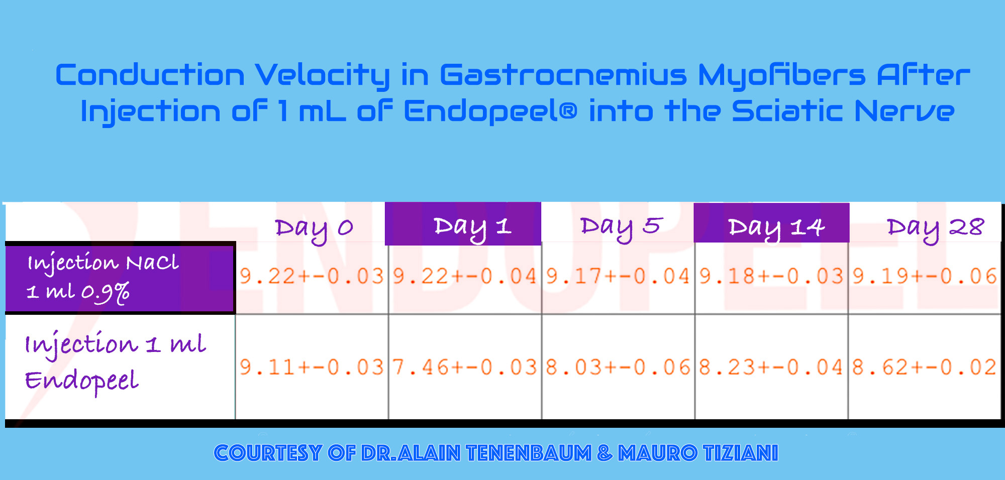
Conduction velocity observations related to Endopeel functional myostructural support
Figure 6. Experimental observations related to conduction velocity in treated tissues, suggesting that Endopeel® may influence not only structural support but also functional behavior within selected myofibers and adjacent support planes.

Conduction Velocity and Functional Myostructural Support

Another important area of investigation concerns conduction-related observations within treated myofibers and adjacent support structures. These findings are consistent with the idea that Endopeel® may influence not only tissue consistency but also functional behavior in selected muscular territories.


This is one of the reasons the technique has been positioned as a method of functional myoplasty or myopexy. The objective is not to artificially occupy space, but to help restore support, tension balance and contour through selective structural action.


  • Functional tissue support
  • Possible interaction with conduction behavior
  • Reinforcement of weak structural planes
  • Improved contour through support, not overfilling

Vascular and Periorbital Effects

In selected indications such as dark circles, the visible improvement may involve more than simple camouflage. Better support of the local tissue environment may influence the appearance of vascular show-through, shadowing and periorbital depression.


Periorbital dark circles are known to have a multifactorial origin including vascular show-through, tear trough depression, shadowing effects and, in some cases, pigmentary components. Structural support of the periorbital tissue may therefore modify the optical appearance of the region by reducing shadowing and improving light reflection across the lower eyelid contour.


The effect on vessels should therefore be interpreted cautiously and in context: not as a classic vascular treatment, but as a structural intervention that can modify how the overlying tissues behave and how vascular coloration becomes clinically visible.

Periorbital implication: improvement may result from better support, reduced transparency effect and modified light reflection.
Clinical improvement of dark circles
Figure 7. Clinical example of periorbital improvement. Structural support of the local tissue environment may reduce the visible appearance of vascular show-through and shadowing, contributing to a more homogeneous periorbital contour.
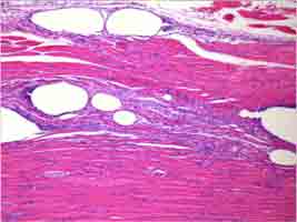
Histological section showing vacuolization phenomena after Endopeel injection
Figure 8. Histological section illustrating vacuolization phenomena observed in treated tissue planes after Endopeel® injection. Such microstructural changes may reflect localized physicochemical interactions within the tissue environment and a transient rearrangement of tissue architecture.

Histology, Microstructure and Experimental Evidence

Histological observations following Endopeel® injections have revealed areas of localized vacuolization and microstructural modification within the treated tissue planes. These findings suggest that the procedure may induce a physicochemical interaction with the surrounding tissue environment rather than producing a purely superficial cosmetic effect.


The presence of vacuolization may represent a transient reorganization of the microstructure of the tissue. Such changes could influence local mechanical properties, including tension distribution and structural behavior of hypotonic tissue compartments.


When these histological observations are considered together with clinical outcomes, they contribute to a plausible biological framework linking localized tissue interaction, microstructural rearrangement and the functional support effects observed in practice.

Additional microscopic material and detailed histopathological observations are available in the dedicated histology section.


See detailed histopathological material →

Duration of Action

The duration of Endopeel® effects may vary according to the indication, tissue quality, technique and functional loading of the treated area. Clinical and observational data nevertheless suggest that some effects are not merely immediate but may persist over several months depending on the treated anatomy.

Immediate
Structural support effect may appear rapidly after injection.
Short-term
Functional improvement may continue to stabilize during the first weeks.
Mid-term
Several clinical observations indicate persistence over a period of months.
Variable
Duration depends on anatomy, indication, technique and maintenance strategy.
Figure 9. Conceptual timeline illustrating the typical progression of clinical effects observed after Endopeel® treatment. Immediate structural interaction may be followed by functional stabilization and persistence of clinical outcomes over several months depending on the anatomical indication and tissue characteristics.

Why These Mechanisms Matter Clinically

Understanding the mechanisms of action is essential to explain why Endopeel® should not be confused with classic fillers, simple volumizing procedures or purely superficial correction techniques.


The proposed mechanisms suggest a treatment rationale based on structural tissue interaction, functional support and selective reinforcement of hypotonic compartments. This framework helps explain the clinical relevance of the technique in indications characterized by tissue laxity, support failure and biomechanical instability.

Mechanistic synthesis

Taken together, these observations suggest that Endopeel® may act through a combination of localized physicochemical interaction, microstructural tissue reorganization and functional support of hypotonic anatomical compartments.


The integrated framework helps explain the clinical effects observed across different indications while remaining compatible with the experimental and histological findings currently available.

  • Functional myoplasty concept
  • Structural support rather than volumization
  • Selective interaction with hypotonic tissues
  • Coherent mechanistic framework
Figure 10. Conceptual summary of the clinical interpretation of Endopeel® mechanisms of action, integrating physicochemical tissue interaction, microstructural modification and functional support effects observed in clinical practice.
Scientific note

The mechanisms described above represent an interpretative framework based on clinical observations, experimental material and histological findings reported in the Endopeel® literature. As with many minimally invasive techniques, several biological aspects remain the subject of ongoing investigation and should be interpreted within the context of currently available evidence.


Continue reading

Experimental Outline

Experimental Study 

Evaluation of the muscular alterations after the intra muscular application of ENDOPEEL

The authors studied the alteration of the gait and the tissue reaction of the muscle which got an endopeels injection.
We chose to apply endopeel into the pre tibial muscle of rats and study the histological alterations.
The choice of the pre tibial muscle of rats was made because of similarity as weight with the depressor muscle of the corner of the mouth in humans.

Objective : Study the biological phenomenon caused by the endopeels injection into the pre tibial muscle of the Wister Rat and evaluate the alterations in its gait and the histopathological alteration in the applied muscle.

who are they

Authors

  1. Main Authors :  
    -Dr.Alain Tenenbaum
    -Sr.Mauro Tiziani

  2. Tomas Yung Joon Kim
  3. Romulo Mené

Read More about the Main Authors

01

Study
Group

Experimentators : R.Mené,M.Kim,A.Tenenbaum,M.Tiziani

N= 29 Wistar rats
Weight mean : 421g

Origin of animals : Biotério Central of Campus of Ribeirão Preto of São Paulo University
Maintenance : Experimental Pathology Lab of Pathology Department on Medicine School of Ribeirão Preto – USP
Animals keeping : polypropylen boxes
Animals food : basic diet of lab and water
Light conditions : natural light/dark cycles under controlled basic conditions
Experimentators : A.Tenenbaum,R.Mené,M.Kim,M.Tiziani

pathology department

02

Animals Sacrifice & Histologic Samples

Conventional Microscopy

The mice were euthanized in CO2 chambers.

  • Incision of the skin with a blade nº 23
  • Finding and isolation of the pretibial muscle using Metzenbaum scissors
  • Section of tendons with a Mayo scissors.
  • Fixation of Portions of the muscles on sections of cork oak
  • Embedding using 50% formaldehyde solution - 10% buffered and 50% of pure paraffin for conventional optical microscopic study.
  • Staining Sections as routine histological process.
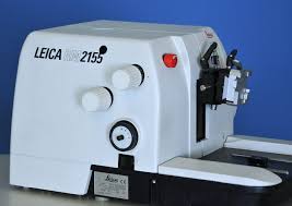
  • Microtome used : Leika RM 2155
  • Coloration of muscle slices with

-hematoxilin-eosin
-Masson´s trichrome
-and red picro-sirius.

  • Histologic examination was realized with a conventional microscope (Olympus BH2)
  • Observation under polarized lampe
  • Photographic documentation was realized with a Leika DMR microscope joined to a digital camera Leika CD 300F and compatible PC.
microtome

03

Material

6 rats/box

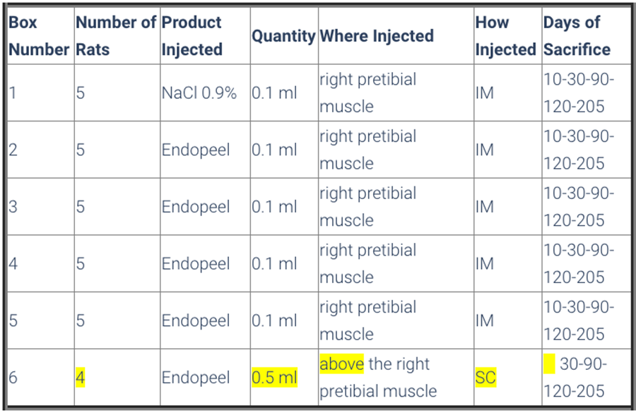
  • Animals were divided in 6 boxes, so that boxes from number 1 to 5 had 5 rats and box number 6 had 4 rats 
  • All animals were submitted to a walking evaluation by footprint impression on special paper , and previous immersion of back legs on a soap solution. 
  • Box 1 had the control group, that received the application of 0,1 ml saline solution ( NaCl 0,9%) on the right pretibial muscle. 
  • The mice of boxes 2 to 5 received 0,1 ml of Endopeels Original Main Product on the same muscle as control group.

  • Box 6 was constituted of 4 rats, and each one received 0,5 ml of Endopeels original main product injected on the subcutaneous layer. 

  • All the animals were submitted to walking evaluation before and after their respective injections.

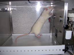
boxes for rats

04

Methods

Animals are sacrified after 10 days-1 month- 3 months-7 months

sacrifice table

Gait Evaluation

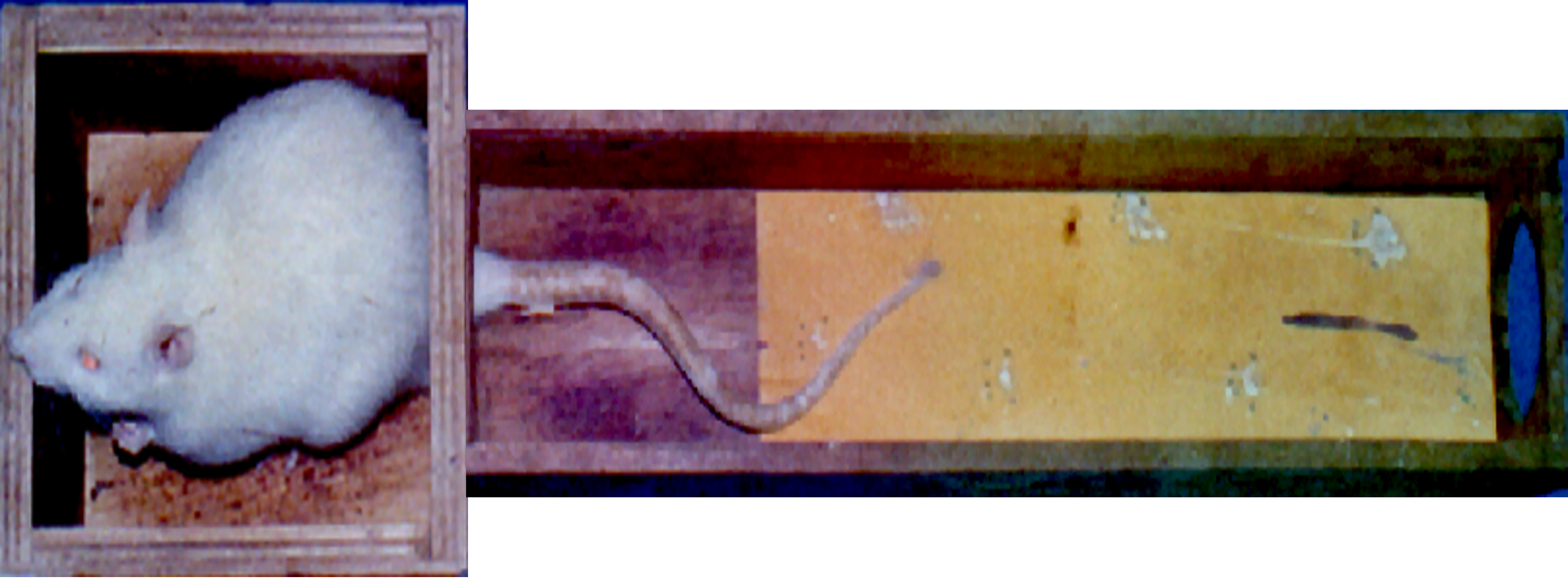
To register the footprints of the rats, strips of paper measuring  43 cm long by 8.7 wide, sizes that are considered as being adequate  for the rats ‘catwalk’ and built as per the De MEDINACELI; FREED; WYATT (1982) drawing. The paper was previously soaked in a solution of bromophenol blue were used.

For the preparation of the paper the bromophenol blue in its anhydrous (Sigma) form was diluted in at 1% in absolute acetone, acquiring an orange color.
The sheets of paper were soaked in this solution and placed in the sun to dry, then they were cut into strips and stored in plastic packages, so as to avoid humidity, which could alter the blue color. 

gait evaluation

The animals were made to walk on the catwalk covered with the previously prepared paper strips, with their hind legs soaked in a solution of neutral detergent, which immediately changes the color of  the footprint from yellow to blue . The footprints remain on the paper strips, which were left to dry for the reading of the necessary measures to calculate the Sciatic Functional Index .
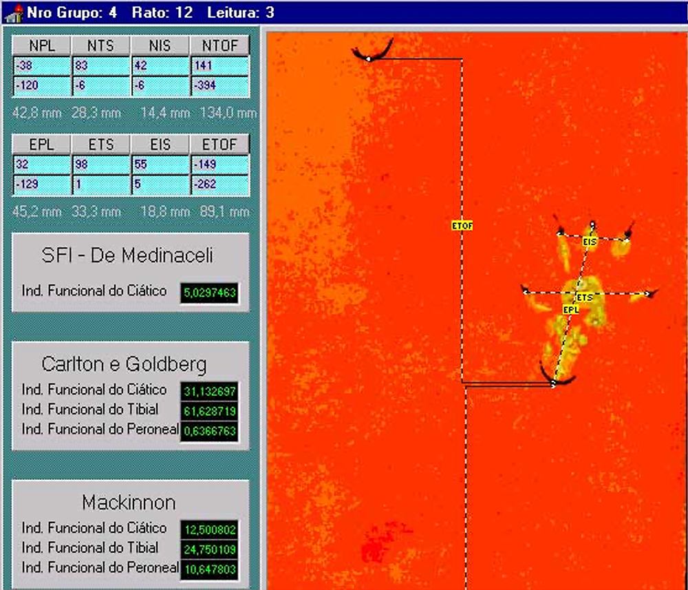
Later these were analyzed by a computer program (software) developed by SELLI (1998) , to obtain the Sciatic Functional Index that was proposed by BAIN; MACKINNON; HUNTER (1989), which takes into account the print length, the total toe spread between the 1st to the 4th toe,  and the intermediate toes which are the 2nd and 3rd.  The footprints of each animal were obtained before the application of endopeel, as a control, and immediately after the endopeel injection, and again, and at periodic intervals, when the animals were sacrificed for the extraction of the anterior tibia muscle  for histological study.

The footprints were digitalized with a scanner and placed into the SELLI program, which allows for the capture, identification and analysis of the images, allowing for the application of other methods of identification of the Sciatic Functional Index (DE MEDINACELI, FREED & WYATT, 1982; DE MEDINACELI, DERENZO & WYATT, 1984; CARLTON & GOLDBERG, 1986), as well as Tibia Functionality indexes and the peroneal nerve (BAIN; MACKINNON & HUNTER, 1989). Once the footprint is shown on the monitor, its parameters are analyzed and measured, which require the placing of the cursor to be clicked in the correct predetermined sequence. The program then calculates and supplies the value of the index, and this is also stored in the computer.

Footprints of the mouse without any injection

Footprints of the mouse after 0.05 ml injection in the right pre tibial muscle

SFI Calculation

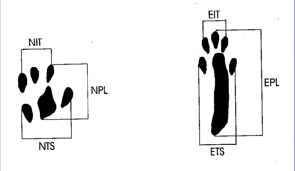
Sciatic Functional Index or SFI Calculation & Measurement

Schematic representation of the measurements realized to calculate the Sciatic Functional Index.

E: Experimental (operated foot);

N: Normal (opposite foot);

TS: Total opening of the toes (1st to 5th);

IT: Opening of the intermediate toes (2nd to 4th);

PL: Length of footstep

SFI endopeel measurements

Program used to measure the Sciatic Functional Index SFI

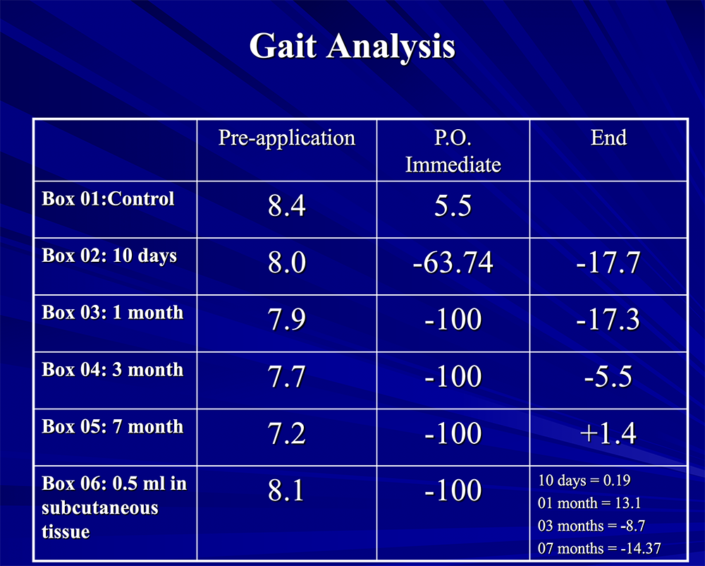
gait analysis

Gait Analysis

Gait Conclusions

  • NaCl 0.9%, 0.1 ml into the pretibial muscle does not alter the gait.

  • Endopeel at 0.1 ml intramuscular pretibial affects the gait accentually,  immediately after the application & moderately after 10 to 30 days.
  • The alteration in the gait caused by Endopeel decreases with time when applied via intramuscular, at this level of concentration.

  • Application into the  subcutaneous tissue is not as efficient as intramuscular.
  • When 0.5 ml is applied into the subcutaneous tissue (pretibial muscle region) there is a progressive loss of gait, probably because of the slow diffusion of Endopeel from the subcutaneous tissue  to the intra muscular one.

Continue reading

Molecular Biology

Molecular Biology of Endopeel
Bioenergetic and Molecular Foundations


Conceptual Origin & Scientific Framework

Bioenergetic Foundations of the Endopeel Model

The molecular biology framework presented on this page reflects the conceptual and scientific work developed by Mauro Tiziani .

His approach integrates bioenergetics, molecular structure, and non-equilibrium biological systems to describe tissue modulation beyond injury-repair models.

This conceptual foundation serves as the scientific backbone for the Endopeel methodology, later translated into clinical protocols and reproducible outcomes.

zaguarino-AI

Core Biological Concept

Bioenergetic Cellular Reorganization

Endopeel is based on a bioregenerative biological model that focuses on restoring intracellular energetic organization rather than inducing tissue injury. Its primary effect is the reduction of oxidative interference affecting DNA-associated macromolecules, allowing intrinsic cellular control mechanisms to resume normal function.

  • Biological Target

    Endopeel acts at the molecular and energetic level of the cell, where oxidative stress disrupts regulatory macromolecular structures involved in gene expression and signal control.

  • Functional Outcome

    By reducing oxidative interference, cellular regulatory pathways regain coherence, leading to improved metabolic coordination without triggering inflammatory repair cascades.

Molecular Structure and Cellular Accessibility

Low Molecular Weight Aromatic System

The molecular design of Endopeel is based on a low molecular weight aromatic structure, selected for optimal cellular accessibility and bioenergetic compatibility.

  • Molecular Weight

    Low molecular weight enables rapid tissue diffusion and direct cellular access without prior enzymatic degradation.

  • Cellular Entry

    The molecule can penetrate the cell through ionic channels and membrane-associated pathways, avoiding metabolic overload.

  • Biological Readability

    Minimal molecular complexity enhances the cell’s ability to recognize and utilize the signal efficiently.

Aromatic Hydrogen Reactivity

Controlled Bioenergetic Instability

A defining characteristic of the aromatic structure used in Endopeel is the relative instability of its hydroxyl-associated hydrogen, conferring a controlled bioenergetic reactivity.

  • Molecular Property

    The unstable hydrogen represents an energetically active site capable of participating in intracellular energy-transfer processes.

  • Biological Advantage

    This reactivity occurs without structural damage, allowing modulation rather than destruction of molecular systems.

Transmembrane Energetics

Electron–Proton Conversion Mechanism

Endopeel’s biological activity involves a bioenergetic interaction at the plasma membrane level, driven by transmembrane electrical potentials.

  • Electrical Potential

    Transmembrane potentials transport electrons across the plasma membrane during normal cellular activity.

  • Molecular Interaction

    These electrons interact with the aromatic structure, preferentially targeting the unstable hydrogen.

  • Proton Generation

    The hydrogen is converted into a proton (H⁺), representing a localized and efficient energetic transformation.

Intracellular Proton Dynamics

Macromolecular Signal Modulation<

The generated protons migrate toward the intracellular environment, where they interact with macromolecular assemblies involved in signaling and metabolic regulation.

  • Macromolecular Effects

    Proton interaction modifies the conformational state of proteins and signaling complexes, improving their functional alignment.

  • Oxidative Neutralization

    These interactions contribute to the hydrolysis and neutralization of oxidative factors that impair molecular communication.

Entropy, Aging, and Metabolic Efficiency

Restoring Energetic Order

entropy

From a molecular biology perspective, tissue aging is associated with increasing biological entropy and loss of metabolic directionality.

  • Aging and Entropy

    Energy dispersion and inefficient coupling between energy and biological work characterize aged tissues.

  • Endopeel Action

    Endopeel reduces local entropy by simplifying energetic inputs rather than increasing molecular complexity.

  • Functional Result

    improved energetic order enhances cellular responsiveness and metabolic coherence.

Avoidance of Complex Molecular Systems

Minimizing Energetic Waste

Highly complex or large molecular systems impose significant energetic costs on biological tissues.

  • Limitations of Complexity

    Such systems require fragmentation, dissipate energy as heat, and generate non-functional intermediates.

  • Endopeel Strategy

    Endopeel relies on bioenergetically efficient, low-complexity molecules to minimize metabolic waste.

Reorganization Rather Than Damage

Clinical and Biological Implications

Endopeel does not depend on tissue injury followed by reparative inflammation. Its objective is energetic and metabolic reorganization.

  • Mechanistic Approach

    Restoration of intracellular fluxes and functional gradients without inducing damage.

  • Tissue Applicability

    Effective even in aged or metabolically compromised tissues.

  • Clinical Expression

    Results are reproducible, coherent, and based on metabolic optimization rather than injury-repair cycles.

Endopeel represents a bioenergetically coherent approach to tissue modulation, where therapeutic efficacy is achieved by restoring metabolic order rather than increasing molecular complexity.

Conceptual Synthesis

Endopeel is not defined by a single mechanism, but by a coherent bioenergetic framework.

By reducing molecular complexity and restoring metabolic order, it enables biological responses without relying on injury-based repair models.

This conceptual foundation underlies all clinical applications of the Endopeel methodology.

Continue reading

Physiology

Experimental Evidence of Functional Muscle Modulation

Endopeel Physiology

This page presents the physiologic foundation of Endopeel® based on experimental muscle contraction studies conducted at Akita University School of Medicine in collaboration with the Kyoto Pathological Research Institute (2006–2007).

Endopeel® physiology experimental muscle contraction analysis

Data shown below are presented as physiologic tracings and comparative stimulation tests (electrical versus chemical), followed by the observed response after sequential Endopeel® injections.

Abstract

Physiologic Summary

This experimental physiologic study evaluated the effects of localized Endopeel® injection on muscle contraction amplitude and intrinsic muscle tone. Controlled measurements were performed using electrical and chemical stimulation protocols.

Results demonstrate reversible modulation of muscle contractility without neuromuscular blockade, denervation, or muscular atrophy. These findings support the classification of Endopeel® as a functional myomodulatory technique rather than a paralytic intervention.

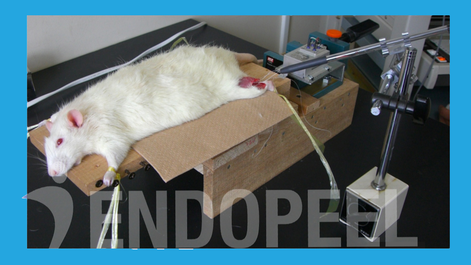
Experimental model used to evaluate myotension (intrinsic muscle tone) and functional muscle behavior following Endopeel® injection
Figure 1. Experimental model used to record baseline tone and functional muscle response (myotension analysis).
Experimental Study

Akita University Physiologic Study

This experimental work (2006–2007) was conducted at Akita University School of Medicine in collaboration with the Kyoto Pathological Research Institute. The objective was to document changes in intrinsic muscle tone (myotension) and the resulting functional behavior of muscle tissue following localized Endopeel® administration.

Observed variations in apparent contraction amplitude are interpreted as a biomechanical consequence of increased tone and structural containment (myoplasty), rather than neuromuscular blockade. In clinical terms, this supports a functional triad: myotension (tone increase), myopexy (functional repositioning), and myoplasty (tissue tightening / confinement).

Authors
  • Prof. Dr. Hirotaro Fukuoka
  • Dr. Alain Tenenbaum, MD, PhD (Hon.)
  • Mr. Mauro Tiziani, Molecular Biologist; Red Cross Surgical Assistant (RCSA)
Baseline Recording

Baseline Muscle Tone and Functional Capacity

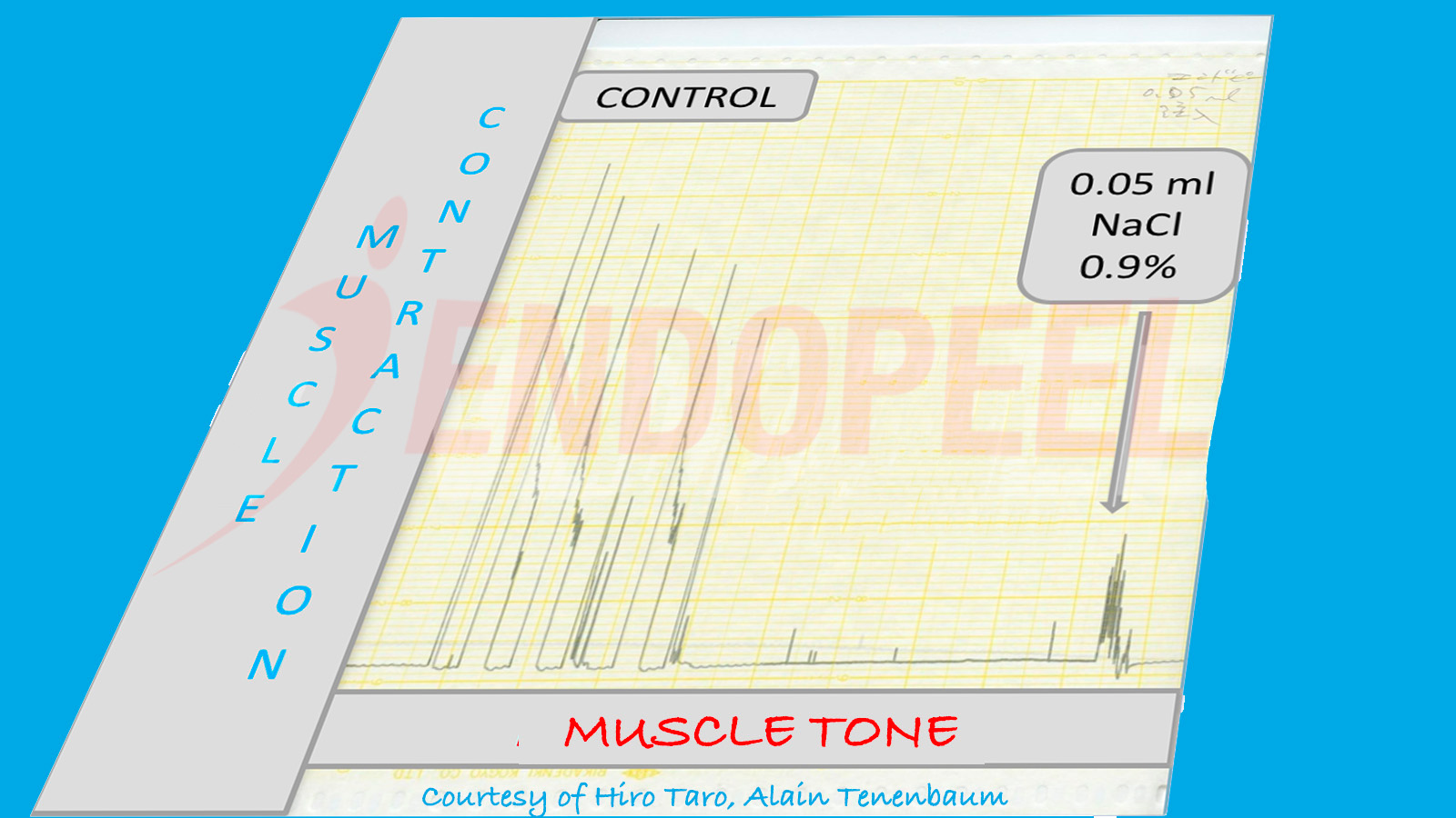
Prior to Endopeel® administration, baseline recordings were obtained to characterize the physiologic state of the muscle: resting tone (myotension) and overall functional responsiveness under standardized stimulation.

At baseline, the muscle demonstrated normal resting tonicity and unrestricted range of motion. This reference phase is essential, because subsequent reductions in apparent contraction amplitude can be interpreted correctly as a mechanical consequence of increased tone and structural confinement (myoplasty), rather than loss of neuromuscular function.

  • Baseline myotension: physiologic resting tone
  • Functional capacity: preserved responsiveness and movement
  • Interpretation framework: amplitude changes ≠ paralysis
Baseline gastrocnemius tracing illustrating physiologic resting tone (myotension) and normal functional response prior to Endopeel® injection
Figure 2. Baseline tracing: physiologic myotension and preserved functional responsiveness prior to injection.
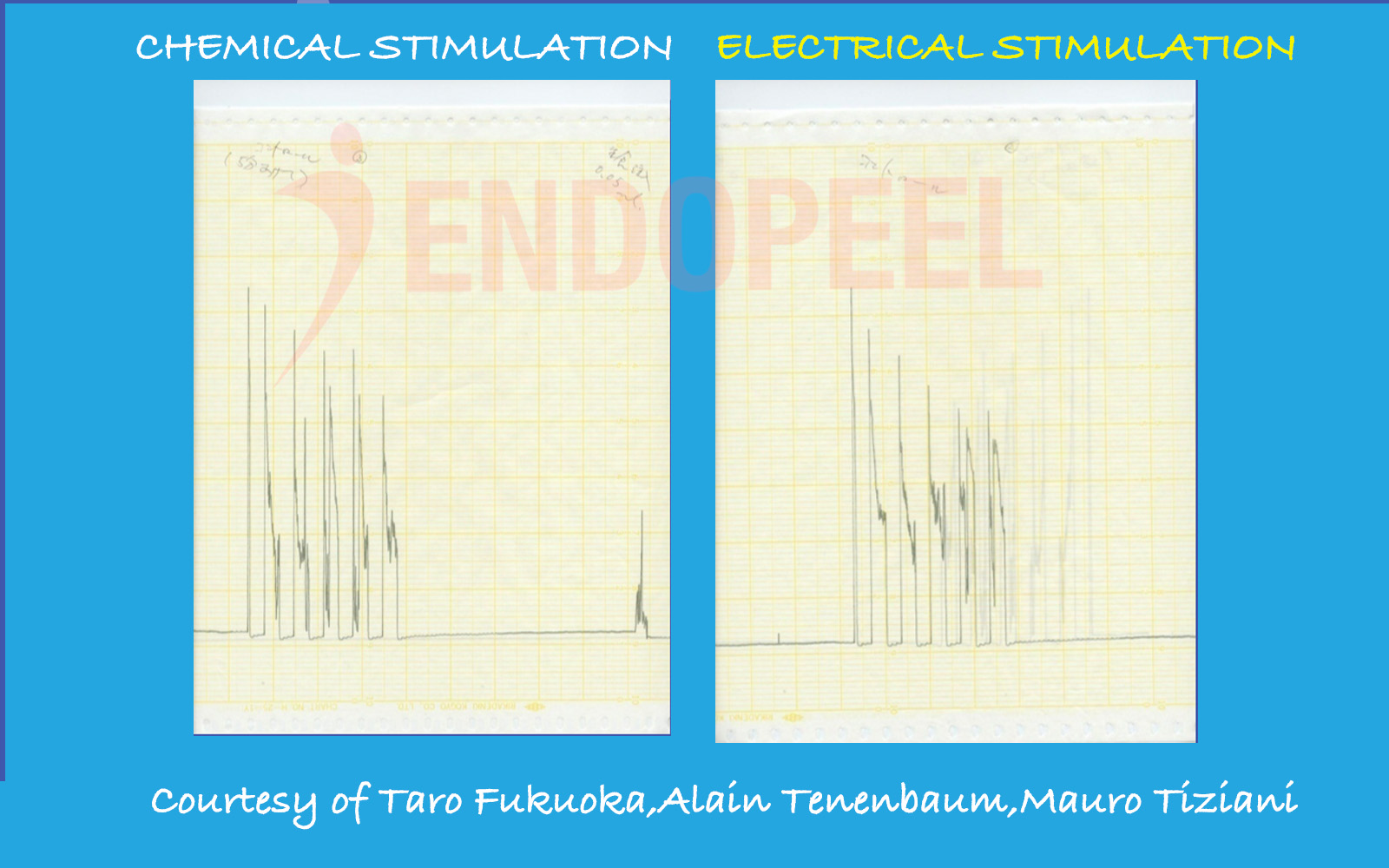
Comparison of electrical stimulation versus chemical stimulation with NaCl 0.9% demonstrating preserved muscle responsiveness prior to Endopeel® injection
Figure 3. Electrical stimulation versus chemical stimulation (NaCl 0.9%): baseline muscle responsiveness prior to Endopeel®.
Pre-Injection Control Phase

Electrical vs Chemical Stimulation (NaCl 0.9%)

Before Endopeel® administration, the muscle was evaluated under two controlled activation methods: electrical stimulation and chemical stimulation with NaCl 0.9%.

The purpose of this comparison is to establish baseline functional integrity: the muscle remains responsive and viable, with physiologic resting tone (myotension) and reproducible activation patterns under both stimulation modalities.

  • Control stimulus: NaCl 0.9% (chemical)
  • Functional endpoint: preserved muscle responsiveness
  • Baseline framework: tone (myotension) and viability established prior to Endopeel®
Early Physiologic Response

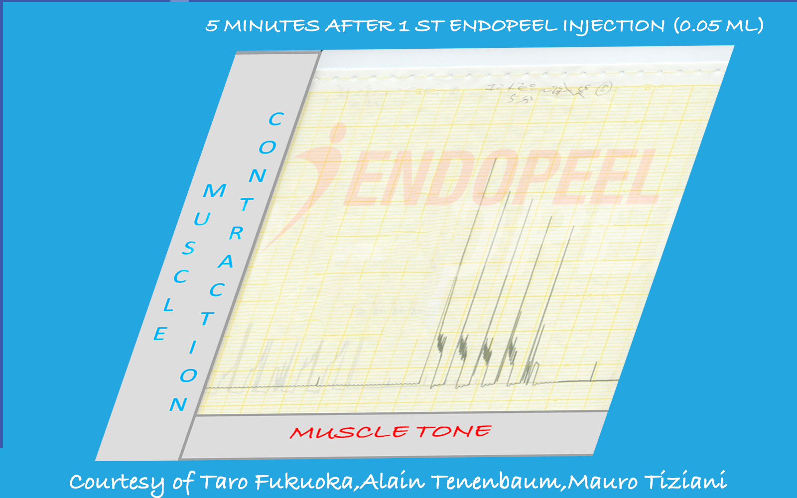
5 Minutes After First Endopeel® Injection (0.05 mL – Single Point)

In this experimental phase, only 0.05 mL was injected at a single intramuscular point. This volume is deliberately minimal and primarily intended to observe early physiologic changes.

At this dose and single-point distribution, measurable increase in global muscle tone (myotension) may remain subtle and not yet produce a visible structural effect.

  • Injected volume lower than typical clinical application
  • Single injection point only
  • Dose-dependent effect expected

Clinically, structural myotension effects are achieved either through multiple distributed injections (0.05–0.1 mL per point) or, when targeting a single confined area such as a trigger zone, through higher localized volume (up to approximately 1 mL).

Muscle tracing five minutes after first 0.05 mL Endopeel® injection at a single point
Figure 4. Early physiologic response after minimal single-point injection (0.05 mL).
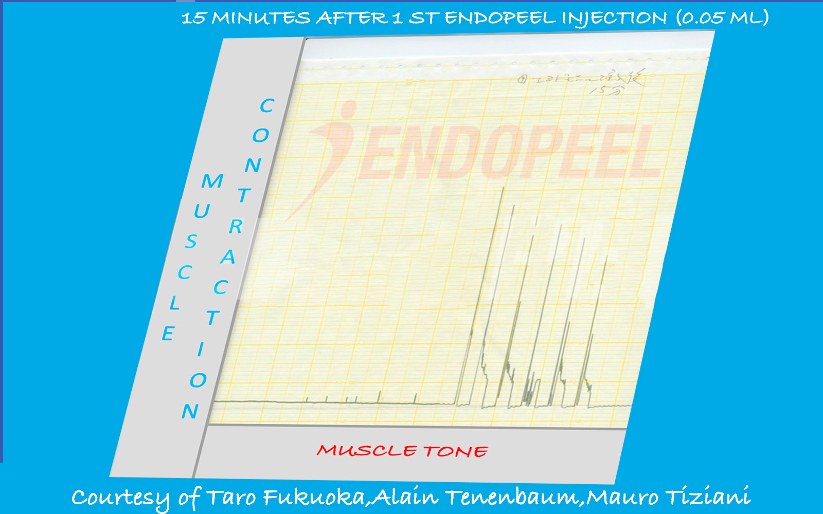
Muscle tracing fifteen minutes after Endopeel® injection demonstrating biomechanical confinement with preserved vitality
Figure 5. Functional adjustment within a progressively confined structural environment.
Structural Adaptation Phase

15 Minutes Post Injection: Myotension and Myoplasty

Fifteen minutes after injection, intrinsic muscle tone (myotension) remains elevated. The muscle progressively adapts to a more compact structural configuration.

Apparent reduction in contraction amplitude at this stage does not indicate neuromuscular inhibition. Instead, it reflects mechanical confinement within a tightened structural envelope — the beginning of the myoplasty effect.

Biomechanical analogy: A muscle operating freely within a large anatomical space can exhibit full excursion. When the same muscle is structurally confined within a smaller environment, its movement remains possible but its amplitude is naturally reduced — without paralysis and without loss of vitality.

  • Myotension remains increased
  • Functional movement preserved
  • Amplitude adjustment is biomechanical, not neurotoxic
Reinforcement Phase

Second Injection: Myotension Reinforcement and Myopexy

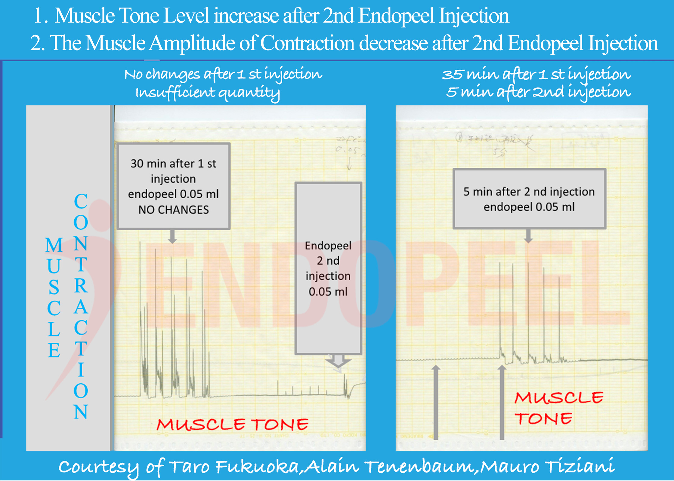
Following the second localized Endopeel® injection, intrinsic muscle tone demonstrates further reinforcement. The structural tightening effect becomes more pronounced.

This cumulative increase in myotension promotes functional repositioning of the muscle within its anatomical compartment — the basis of the myopexy effect.

As structural confinement intensifies (myoplasty), movement range adapts naturally. The muscle remains viable and contractile, yet operates within a more compact and stabilized framework.

  • Reinforced intrinsic tone (myotension)
  • Functional repositioning (myopexy)
  • Structural containment (myoplasty)
Muscle tracing following second Endopeel® injection demonstrating reinforced myotension and structural stabilization
Figure 6. Reinforced myotension and progressive structural stabilization after second injection.
Conclusion

Functional Myotension Without Paralysis

The experimental findings demonstrate that Endopeel® does not induce neuromuscular blockade. Instead, its primary mechanism is an increase in intrinsic muscle tone — myotension.

Progressive tone reinforcement produces structural tightening of the muscular compartment (myoplasty), leading to functional repositioning of the muscle within its anatomical environment (myopexy).

Apparent reductions in contraction amplitude observed in the experimental model reflect biomechanical confinement, not paralysis. Muscle vitality and functional capacity remain preserved.

These experimental observations describe physiologic behavior and should be interpreted within the context of the experimental model.

FAQ

Continue reading